Fact Friday
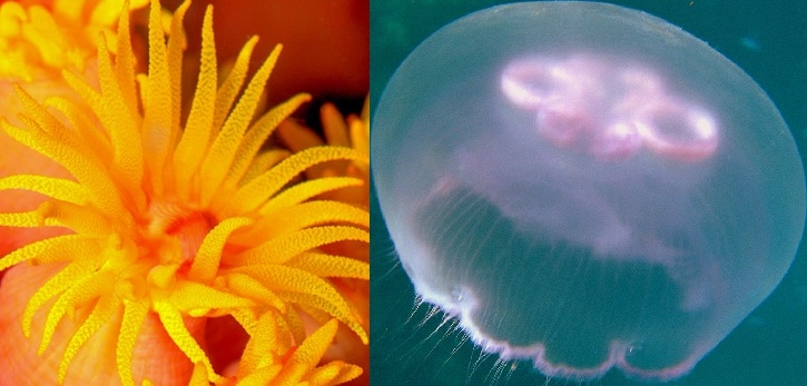
Some cnidarians switch between a polyp and a medusa body form. Corals and anemones only have the polyp stage. Jellyfish, as we think of them, are in the medusa stage, but they have a polyp stage when they are juveniles. Hydrozoans have the opposite: their adult form is a polyp, but they have a juvenile medusa stage.
Photo Credit: KSLOF, Rob Martimbeault


Background Information
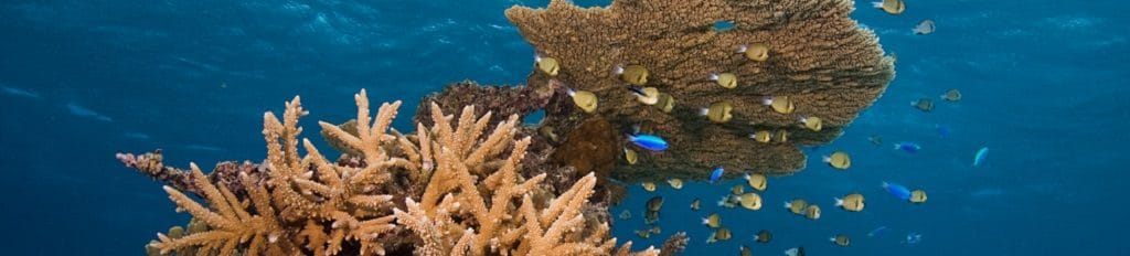
HOW DO CORALS GROW?
In Unit 5: Coral Reproduction, we learned that corals are able to create more coral polyps by reproducing sexually and asexually, but this doesn’t explain how their skeletons are built. Stony corals increase the size of their skeletons by gradually depositing layers of calcium carbonate (CaCO3). This process is known as calcification. Calcification is the rate at which corals create their hard skeletons by absorbing calcium from seawater. In this unit, we will learn how corals grow and the shapes that they form.
How do corals grow? In colonial corals, each polyp contributes to the growth of the coral (figure 9-1). The polyp lifts itself up out of the cup-like skeleton, called the corallite. Then it deposits calcium carbonate in the space below. The polyp secretes this mineral from the basal plate. This layer of calcium carbonate allows the coral skeleton to grow upward. For more information about the structures of a coral polyp see Unit 3: Coral Anatomy.
Fun fact
X

Fun Fact
Do you know that coral exoskeletons can be used as a bone graft? The internal structure and composition of a coral’s skeleton is compatible to that of our own bones. These grafts can be used when someone has a severe bone fracture or when small pieces of bone are missing. When this occurs, the coral exoskeleton is transplanted in the fractured or missing area allowing the bone to grow and repair itself.
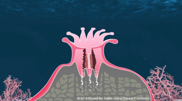
Figure 9-1. Cross section of a coral polyp – the polyp is lifting out of the corallite and depositing calcium carbonate in the space below.
Polyps create calcium carbonate through a chemical reaction. This reaction takes place inside the tissues of the coral polyp. Corals begin this process by pulling calcium and carbonate from the surrounding seawater. Here is the calcification chemical equation:
2HCO3– + Ca2+ → CaCO3 + CO2 + H2O
Bicarbonate + Calcium → Calcium carbonate + carbon dioxide + water
Scientists are not entirely sure how some of this process occurs.

GROWTH RATES
Some coral species have faster growth rates than others. Their growth rate depends on their rate of calcification; the greater the calcification, the greater the growth rate.
Let’s take a look at two different species of coral in the northeastern Caribbean, Acropora palmata or elkhorn coral (figure 9-2a) is a very important reef-building coral in the Caribbean and Florida. It has a fast growth rate of up to 4 inches (10 cm) per year (Gladfelter et al. 1978). Other corals are slower growing, such as the boulder star coral, Montastraea annularis (figure 9-2b). This coral only grows about 0.3 inches (7.6 mm) per year (Gladfelter et al. 1978).
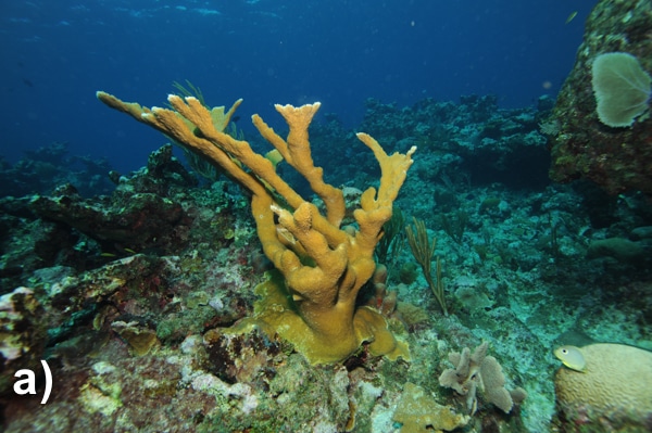

Figure 9-2. a) Acropora palmata; b) Montastrea annularis
Photo Credits: a) Phil Renaud; b) Ken Marks
Corals require a lot of energy for calcification to take place. The energy mainly comes from the photosynthesizing zooxanthellae. Corals photosynthesize (see Unit 4: Coral Feeding) more during the day when there is greater light availability. Research shows that corals also have higher levels of calcification during the day than at night when it’s dark (Kawaguti & Sakumoto 1948; Gattuso et al. 1999). Since photosynthetic activity affects the amount of calcification in corals, this process is often referred to as light-enhanced calcification.
Growth rates of corals are also determined by environmental factors (see Unit 8: Environmental Conditions). Here are some of the main factors that influence photosynthesis and the calcification process.
- Temperature: Studies show that corals calcify best when the temperature is stable and at an optimal range (Clausen & Roth 1975; Coles & Jokiel 1978; Marshall & Clode 2004). This range is different depending on the coral species and their location, but most prefer somewhere between 60.8–86°F (16–30°C).
- Calcium carbonate saturation: The amount of calcium and carbonate in the ocean is called calcium carbonate saturation. Corals need calcium and carbonate to create their skeletons. A low calcium carbonate saturation inhibits calcification.
- Turbidity: Corals receive less sunlight when turbidity is high because the particles block the sunlight. This inhibits photosynthesis from taking place.
- Sedimentation: Sedimentation takes place when sediment particles are suspended in the water. Corals that are smothered by sediment, receive less sunlight. This also inhibits photosynthesis from taking place.
- Salinity: Corals calcify at an optimal salinity range. The more stable the salinity, the more likely corals will calcify. Again, this varies per coral species, but is usually between 23-42 ppt.
- pH: As the pH of seawater becomes more acidic, it inhibits calcification. We will learn more about this process when we discuss ocean acidification in Unit 19: Threats (coming soon).
- Currents: When water flow is greater, corals are able to increase their calcification rate; however, too great of water movement can inhibit coral growth. Moderate water flow increases the exchange of gases (CO2 and O2), inorganic nutrients, and food availability. Corals need gases and inorganic nutrients to photosynthesize. As we just learned, the more corals photosynthesize, the greater their calcification rates. Moderate water flow also can allow for a greater abundance of food in the water. When corals are able to catch more food, they are able to gain more energy allowing corals to be able to increase their ability to create their stony skeletons. For more on currents see Unit 7:Distribution.
GROWTH FORMS I
Why would scientists need to know a coral’s growth form? Corals are very difficult to identify. Using their growth forms can help scientists identify corals.
As corals grow, they begin to take certain shapes or forms known as growth forms. Each stony coral can be classified by one of these growth forms. Additionally, there are some growth forms that have sub-categories. For simplification, we will learn about the main growth forms.
Branching (figure 9-3): Just like a tree, these corals form branches. These branches can also grow branches called secondary branches. They are not the sturdiest of corals and their branches can break off, but they can use this to their advantage. Branching corals can use fragmentation, a type of asexual reproduction, to create new coral colonies (see Unit 5: Coral Reproduction). These corals typically have faster growth rates than other growth forms.
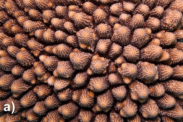
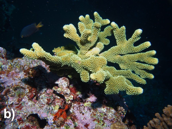
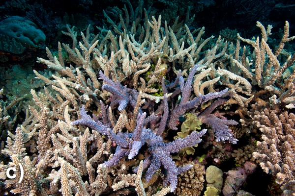
Figure 9-3. a) Acropora sp.; b) Pocillopora sp.; c) Acropora sp.
Photo Credits: a) and c) Ken Marks; b) Andrew Bruckner
Columnar (figure 9-4): These corals are cylindrical in shape and they look like fingers. Columnar corals grow upwards. They differ from branching corals in that they do not have secondary branches.
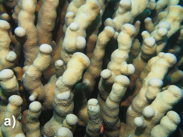

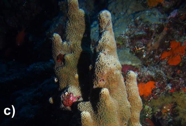
Figure 9-4. a) Pavona clavus; b) Pavona clavus; c) Coscinaraea columna
Photo Credit: Andrew Bruckner
Encrusting (figure 9-5): This form typically adheres to rocky substrates. Encrusting corals do not grow upwards, but instead they grow outward, covering the rocky surface. These corals are able to withstand a great amount of wave action.
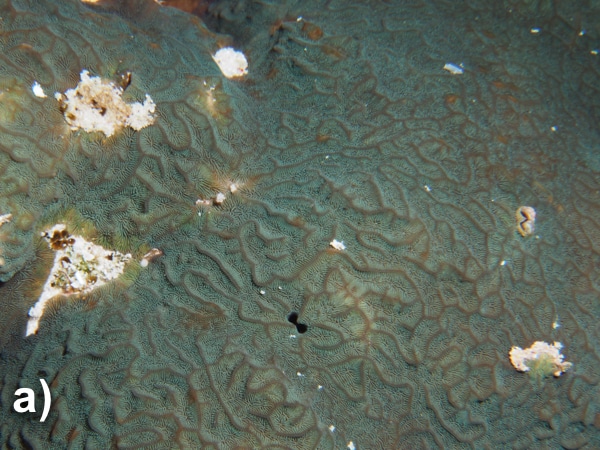
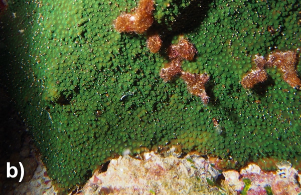
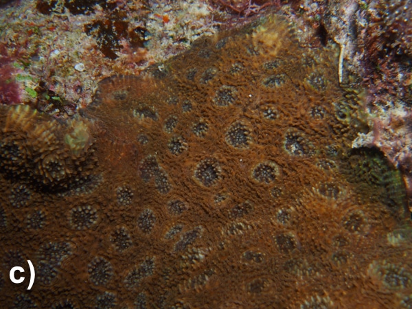
Figure 9-5. a) Pavona varians; b) Cyphastrea sp.; c) Acanthastrea echinata
Photo Credit: Andrew Bruckner
Foliose (figure 9-6): Corals form growth patterns like layered petals in an open flower. These folds increase surface area in order to obtain the maximum amount of sunlight so that their zooxanthellae can photosynthesize.
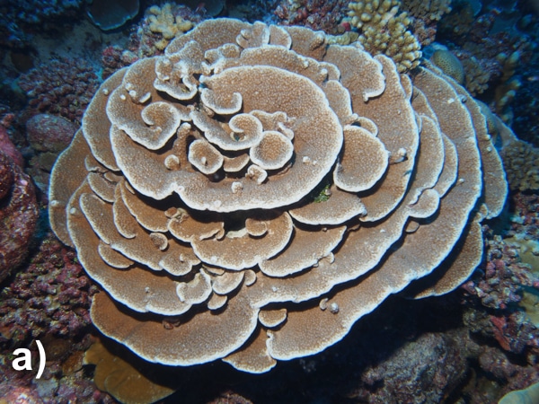
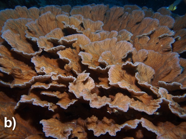
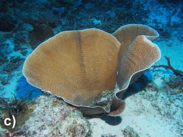
Figure 9-6. a) Astreopora randalli; b) Merulina ampliata; c) Turbinaria sp.
Photo Credit: Andrew Bruckner
GROWTH FORMS II
Free-living (figure 9-7): Not all corals are colonial; some are solitary. Free-living corals are individual coral polyps. They are typically round, oval, or oblong. Some are free-living, while others are attached to a substrate.
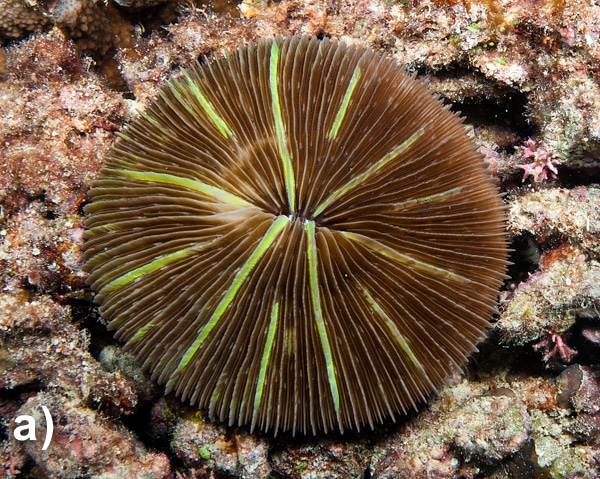
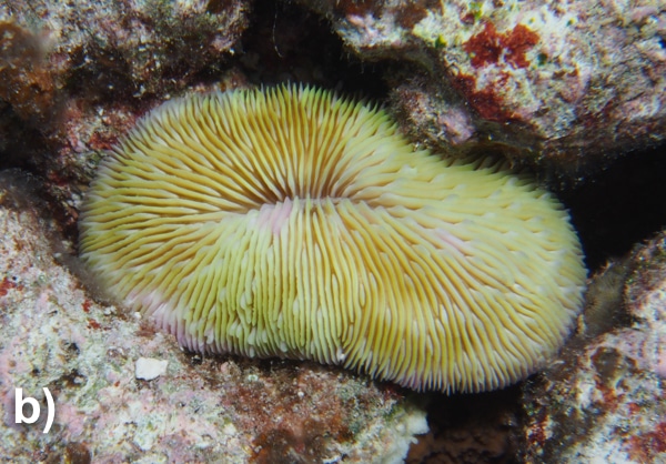

Figure 9-7. a) Fungia sp.; b) Fungia sp.; c) Herpolitha limax
Photo Credits: a) Ken Marks; b) and c) Andrew Bruckner
Massive (figure 9-8): These corals look like domed boulders. They are slow growing and sturdy. They can withstand strong wave action.
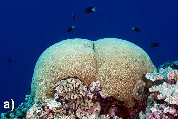
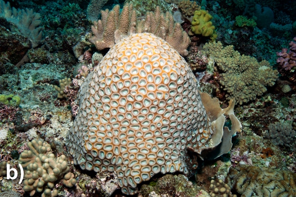
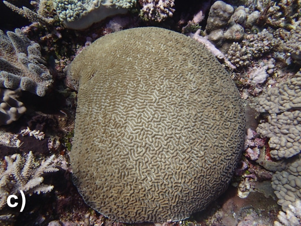
Figure 9-8. a) Astreopora myriophthalma; b) Favites sp.; c) Platygyra sinensis
Photo Credits: a) and b) Ken Marks; c) Andrew Bruckner
Phaceloid (figure 9-9): Each corallite has an individual wall. The corallite is tubular in shape and extends from a common base.
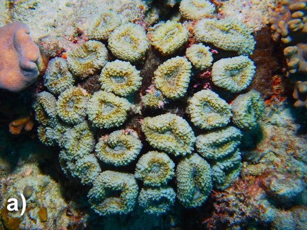


Figure 9-9. a) Lobophyllia corymbosa; b) Lobophyllia hemprichii; c) Caulastrea furcate
Photo Credit: Andrew Bruckner
Plating (figure 9-10): They are thin, plate-like corals. They grow horizontally and look like shelves. Often, plating corals are found in deeper parts of the fore reef (see Unit 11: Zonation). Like foliose corals, they grow horizontally, increasing their surface area in order to obtain the maximum amount of sunlight, so that their zooxanthellae can photosynthesize. This plate-like structure also allows these corals to catch food that is drifting down the reef slope.
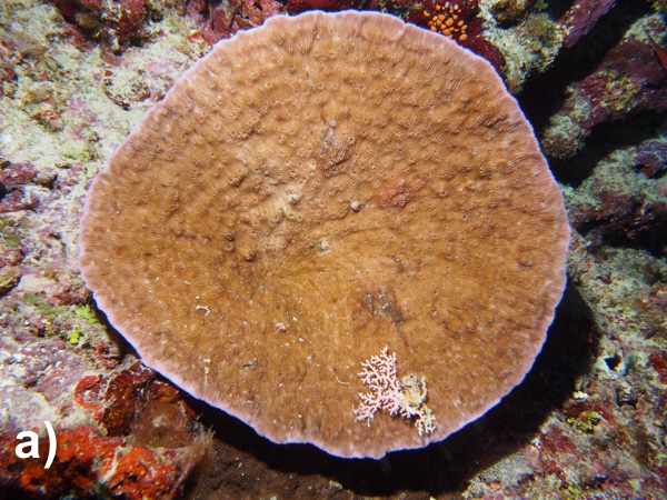

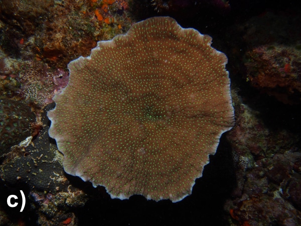
Figure 9-10. a) Leptoseris sp.; b) Mycedium sp.; c) Merulina ampliata
Photo Credit: Andrew Bruckner
CITATIONS
Clausen, C. D. & Roth, A. A. (1975). Effect of temperature and temperature adaptation on calcification rate in the hermatypic coral Pocillopora damicornis. Marine Biology 33: 93-100.
Coles, S. L. & Jokiel, P. L. (1978). Synergistic effects of temperature, salinity and light on hermatypic coral Montipora verrucosai. Marine Biology 49: 187-195.
Gattuso, J., Frankignoulle, M., Bourge, I., Romaine, S., & Buddemeier, R. (1998). Effect of calcium carbonate saturation of seawater on coral calcification. Global and Planetary Change 18: 37-46.
Gladfelter, E. H., Monahan, R. K., & Gladfelter, W. B. (1978). Growth rates of five reef-building corals in the northeastern Caribbean. Bulletin of Marine Science 28: 728-734.
Kawaguti, S. & Sakumoto, D. (1948). The effect of light on the calcium deposition of corals. Bulletin Oceanographic Institute of Taiwan 4: 65-70.
Marshall, A. T. & Clode, P. (2004). Calcification rate and the effect of temperature in a zooxanthellate and azooxanthellate scleractinian reef coral. Coral Reefs 23: 218-224.