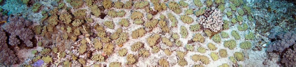
Fact Friday
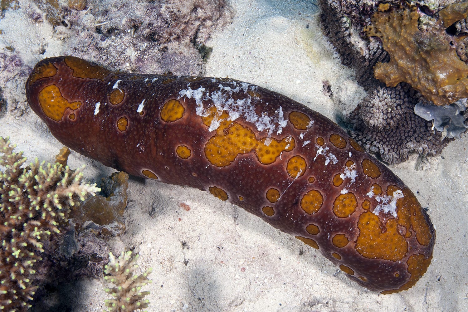
Sea cucumbers are often referred to as “earthworms of the sea.” These strange-looking creatures are extremely beneficial to coral reefs. They use their tentacles to shovel sand, tiny organisms, and detritus into their mouths. Similar to earthworms, they provide “fertilizer” or food for other animals. They do so by excreting waste products such as calcium carbonate and ammonia, which provides nutrients for other animals like corals.
Photo Credit: Ken Marks


Background Information
Coral Life Cycle
A life cycle is a series of events in an organism’s life, as it changes from one form to the next, and eventually returns to the starting state, by means of reproduction. In this lesson, we will learn about the cell cycle and the life cycle of corals.
Fun fact
X

Fun Fact
Can you imagine remaining underground for almost 17 years? This is part of the life cycle of the periodical cicada, often referred to as the 17-year cicada, (Magicicada septendecim). These insects emerge from their eggs as antlike nymphs, and then they drop out of the tree where their mothers laid their eggs. The antlike nymphs bury themselves between 2 to 24 inches into the soil and they latch on to the roots of plants with their mouths. They will feed on the fluid of the roots for the next 17 years! After this time, the mature nymphs dig upwards through the soil until they are about one inch from the soil’s surface. When the time is right, the nymphs emerge from the ground, molt into adult cicadas, mate, and then the females deposit their eggs. Then the cycle begins again. After all of those years of waiting, the cicadas only live above ground for a few weeks. That’s a long time to wait!
In Unit 5: Coral Reproduction, we learned that there are two ways that corals undergo sexual reproduction. There are broadcast spawners and brooders. The broadcast spawner’s life cycle is shown in figure 6-1. The numbers referenced below correlate with the numbers on the diagram.
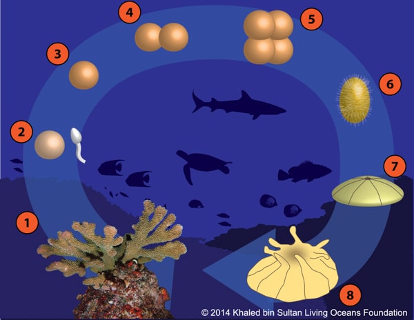
Broadcast Spawners:
- Mature corals use energy to produce gametes. Gametes are produced through meiosis (see Meiosis Section).
- When the environmental conditions are right, corals release their gametes (egg and sperm) into the water column. The gametes float to the surface because they are positively bouyant. External fertilization takes place between male and female gametes (see Unit 5: Coral Reproduction).
- The egg is now fertilized and begins to develop. The fertilized egg is called a zygote. During this phase, the zygote drifts in the current.
- The zygote continues to drift and begins to go through cell division. This type of cell division is known as mitosis (see Mitosis Section). After the first division, there are now two cells.
- Each of these two cells undergo mitosis, creating 4 new cells. These cells continue to go through mitosis, dividing over and over again, creating an embryo.
- A larva forms called a planula. The planula is a type of zooplankton. In this case, it’s a coral plankton. The planula is able to maneuver by the cilia that covers its body. These planulae are microscopic and although they have the ability to move, they are not strong enough to swim against the current. During this phase, the planula is looking for a suitable solid substrate to settle on.
Fun fact
X
Fun Fact
Can corals sense different odors? Scientists believe that in some instances, coral larvae may be attracted by the pleasant odors produced by healthy coral reefs and deterred by the smell of unhealthy reefs that are covered in seaweed. Coral larvae can’t survive on a reef where there is a lot of seaweed because the seaweed will overgrow the larvae and kill it. The larvae will die because the zooxanthellae living inside the coral will not receive the light they require in order to survive. By settling on healthy coral reefs, it gives the larvae a better chance of survival into adulthood.
Source: Dixson, D.L. Abrego, D., & Hay, M.E. 2014. Chemically mediated behavior of recruiting corals and fishes: A tipping point that may limit reef recovery. Science 345(6199): 892-897.
- The planula settles on a hard substrate and begins undergoing metamorphosis. Metamorphosis occurs when an organism develops from a juvenile to an adult.
- The juvenile polyp begins to lay down a calcium carbonate (CaCO3) corallite. If the coral is colonial, then the polyp will go through asexual reproduction to form more polyps, expanding the size of the coral colony and increasing the number of coral polyps. When the adult polyps become sexually mature, the life cycle will begin all over again (steps 1-8).
Fun fact
X
Fun Fact
Some coral larvae have the ability to undergo reverse metamorphosis. What does this mean? It means that the planula larva settles as usual. They lay down a calcium carbonate skeleton attaching to a substrate. Then they metamorphose into a polyp with a mouth, tentacles, and mesenteries. If the polyp becomes stressed within a certain period of time, it can undergo reverse metamorphosis. The polyp is able to reabsorb the calcium carbonate skeleton and reform into a planula larva. This “secondary larva” can resettle and metamorphose into a coral polyp once again.
Source: Richmond, R.H. 1985. Reversible metamorphosis in coral planula larvae. Marine Ecology Progress Series 22: 181-185.
Brooders:
- Corals expend more energy to create a fully developed planula.
a. Brooding corals release only negatively buoyant (when an object does not float in a liquid) sperm into the water column.
b. The sperm finds unfertilized eggs in an egg-carrying coral. Fertilization takes place within the coral.
c. The fertilized egg (zygote) undergoes mitotic cell division (see Mitosis Section) forming an embryo.
d. The embryo grows into the planula within the brooding coral. - The planula is released from the coral polyp. Steps 6-8 are the same as broadcast brooders. See the steps above.

Cell CYCLE
All life begins with a single cell. Just like living organisms go through a life cycle (which we just saw with corals in the previous section), all cells go through a cell cycle.
How do we go from a single cell to a complex, multicellular organism? The answer is cell division. Cell division is when a parent cell divides into two daughter cells. From one cell, the cell divides, creating two cells. Then the two new cells divide creating four cells, and so on and so on. Our bodies are made of trillions of cells and they are constantly dividing!
Cell division is necessary for life. Not only do cells divide as organisms grow and develop, but more cells are created to repair damaged or old and worn out cells. For instance, if you scrape your knee, you bleed, a scab forms, and eventually new skin replaces the scab. This all occurs because new cells are being created. Your skin cells divide to create more skin cells to replace the ones that were damaged.
Cell theory states that cells can only arise from other living cells. The theory also states that all organisms are composed of one or more cells and cells are the most basic unit of life.
Organisms from the Domain Eukarya have complex cells that divide and replicate in a cell cycle. Eukaryotes have two different cell cycles: meiosis and mitosis. Each of these cell cycles plays a vital role in the larger life cycle of an organism.
As we dive into cell cycles, refer to the Appendix if you need to review the parts of a chromosome or cell.

MITOSIS
The first cell cycle we will look at includes a type of division called mitosis. During this cycle, one parent cell divides to create two identical daughter cells that have their own nucleus and identical chromosomes. During mitosis, organisms have diploid cells (2N), which are cell cycles that have two copies of each chromosome (figure 6-2).
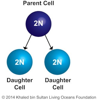
Figure 6-2. Mitosis: cells produced by each division and number of chromosomes
There are two parts that make up this cell cycle – interphase and mitosis. Before cell division begins, the cell is in interphase. Interphase makes up the majority of the cell cycle (figure 6-3). This phase prepares the cell for division during mitosis and can be broken into three phases: the growth one phase or Gap 1 (G1), followed by Synthesis (S), and then growth two phase or Gap 2 (G2).
| Phase | What happens? |
| G1 | The cell grows, doubling in mass and organelles in preparation for cell division. |
| S | DNA is replicated. Each chromosome gains an identical copy. |
| G2 | Microtubules are replicated. They help the cell separate chromosomes during mitosis. The cell is ready to enter mitosis. |
The mitotic phase, sometimes referred to as the M phase, only lasts for a short period of time. During this phase, the chromosomes separate and undergo cytokinesis. Cytokinesis is the division of the parent cell into two daughter cells (figure 6-2). There are several different mitotic phases (figure 6-3).
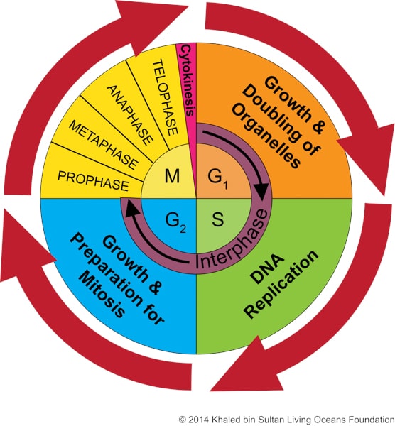
Figure 6-3. Mitosis is only the M part. The cell cycle is continuous, which is illustrated by the red arrows.
We can remember the acronym PMAT to help us remember the order of cell division.
Prophase
Metaphase
Anaphase
Telophase

Mitosis Phases
The four phases of mitotic division are: prophase, metaphase, anaphase, and telophase, which is then followed by cytokinesis.
| Phase | What happens? | Diagrams |
| Prophase | • The chromosomes condense (shorten).
• Centrosomes begin moving toward opposite poles. |
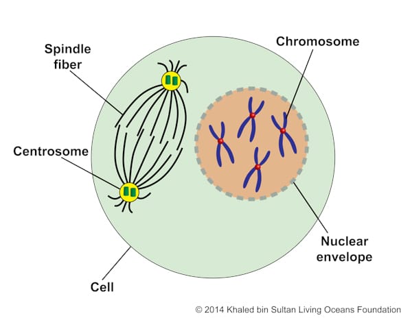 Figure 6-4 |
| Prometaphase
NOTE: There are many steps that take place during prophase and metaphase that scientists often add this additional phase. |
• The nuclear envelope disappears. The nuclear envelope is a double- layered membrane that surrounds the nucleus, separating the nucleus from the cytoplasm.
• Two kinetochores form at each centromere. • Spindle fibers attach to the kinetochores. • Chromosomes begin migrating toward the center of metaphase plate. |
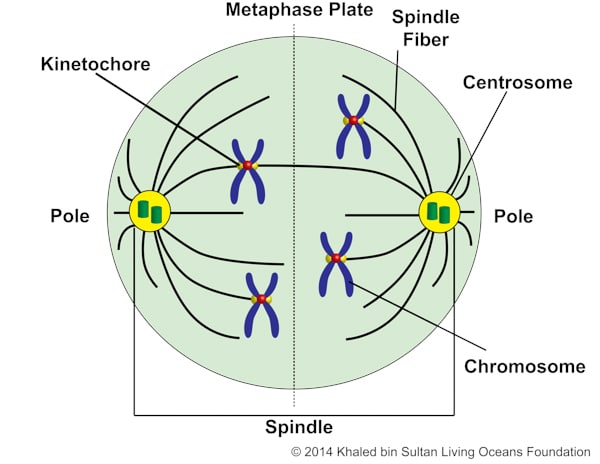 Figure 6-5 |
| Metaphase | • Chromosomes align at the metaphase plate. | 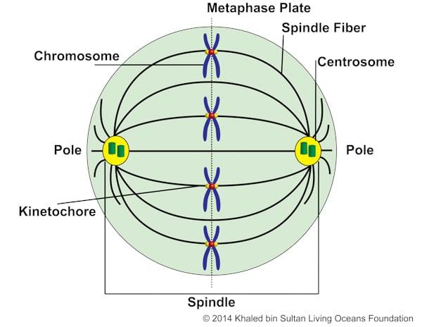 Figure 6-6 |
| Anaphase | • Chromosomes split apart into sister chromatids (identical copies of chromatids).
• The spindle fibers move the chromatids toward opposite poles. • The cell starts to elongate in preparation for division. |
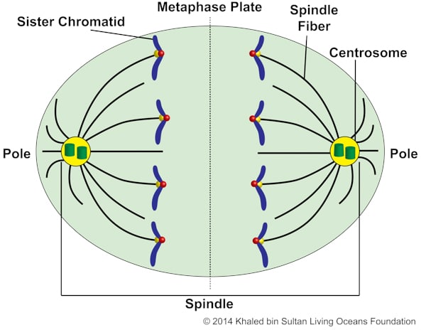 Figure 6-7 |
| Telophase | • In preparation for division, the cell continues to elongate, forming a cleavage furrow (an indentation of the cell’s surface).
• The chromosomes uncoil. • A new nuclear envelope forms. The spindle fibers disappear. |
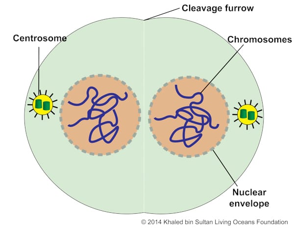 Figure 6-8 |
| Cytokinesis | • The parent cell divides into two daughter cells. After cytokinesis takes place, the cell cycle continues again with the new cells going into interphase. |  Figure 6-9 |

MEIOSIS
All sexually reproductive organisms create mature reproductive sex cells (egg and sperm) called gametes. They create these cells through a process known as meiosis.
Meiosis consists of two different stages – Meiosis I and Meiosis II (figure 6-10). Before meiosis can begin, the parent cell must undergo interphase. In mitosis and meiosis, interphase goes through the same steps. See Mitosis Section for more information about interphase. Note that interphase does not occur between Meiosis I and Meiosis II.
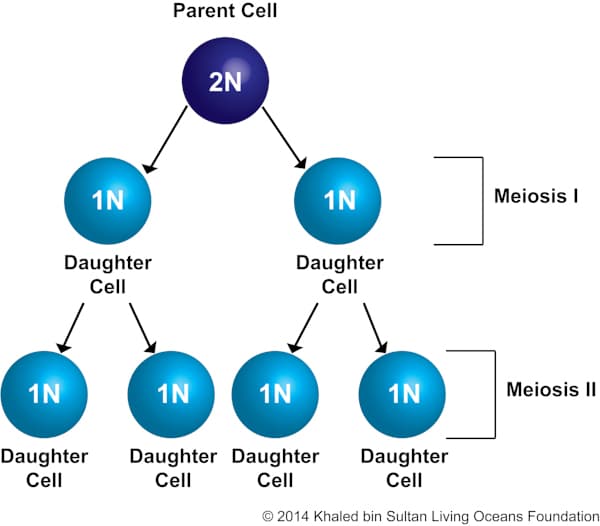
Figure 6-10. Meiosis: cells produced by each division and number of chromosomes
The process of meiosis begins with one parent cell, which is diploid (2N) meaning it has two copies of the chromosomes (figure 6-10). During Meiosis I, cell division takes place creating two daughter cells, which are haploid (1N) meaning that there are half the number of chromosomes of the parent cell (figure 6-10). These two haploid cells then enter Meiosis II and the cells undergo division again, without replication of the DNA. The result of this division is four daughter cells that are each haploid (1N) (figure 6-10).

Meiosis Phases
Meiosis I:
| Phase | What happens? | Diagrams |
| Prophase I | • The chromosomes condense (shorten).
• The nuclear envelope disappears. • Centrosomes move towards opposite poles. • Homologous chromosomes (one paternal and one maternal copy of a chromosome) become paired. From this point on they are known as a tetrad. Each tetrad is composed of four chromatids. • Crossover can take place (figure 6-12). Crossover is the process of exchanging chromosome segments (DNA) between chromosomes. In other words, segments of chromosomes from mom switch spots with the same part of the chromosomes from dad. The area where crossover takes place is called the chiasma. • This is the longest phase of meiosis. |
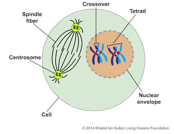 Figure 6-11 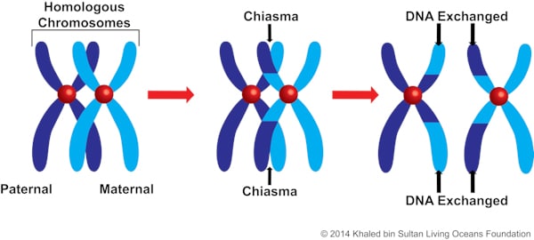 Figure 6-12 |
| Metaphase I | • Spindle fibers attach to the kinetochore.
• Tetrads become aligned at the metaphase plate. |
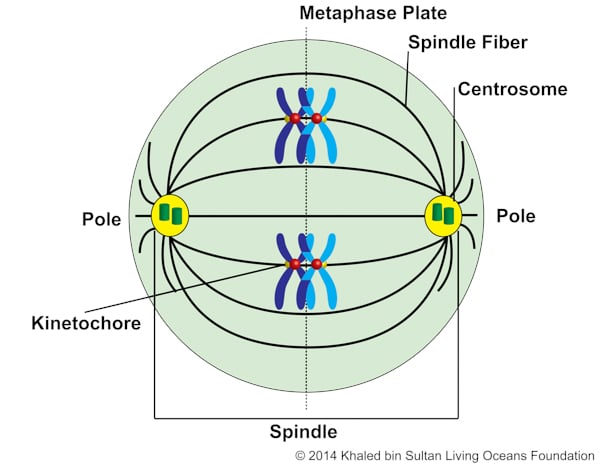 Figure 6-13 |
| Anaphase I | • The spindle fibers split the tetrad and move homologous chromosomes toward opposite poles.
• The cell starts to elongate in preparation for division. |
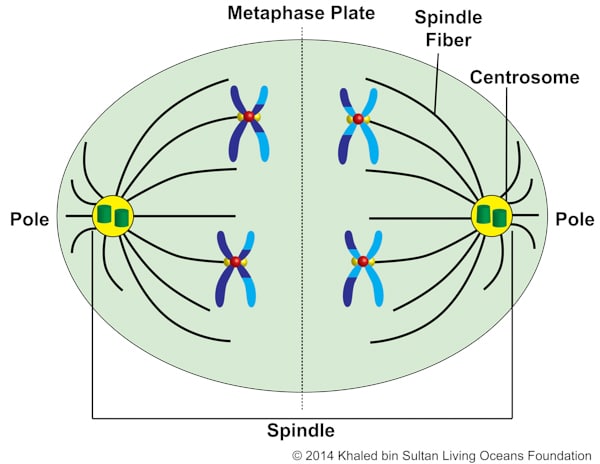 Figure 6-14 |
| Telophase I | • In preparation for division, the cell continues to elongate, forming a cleavage furrow.
• The nuclear envelope reforms around the chromatids. The spindle fibers disappear. |
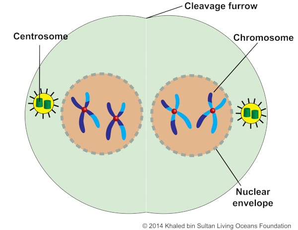 Figure 6-15 |
| Cytokinesis I | • The parent cell divides into two daughter cells. After cytokinesis takes place, the cycle continues. Each cell is now haploid. Figure 6-16 shows the chromosomes uncoiled, which does not always happen during telophase I. | 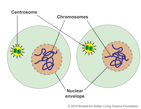 Figure 6-16 |
Meiosis II:
| Phase | What happens? | Diagrams |
| Prophase II | • If the chromosomes have decondensed, then they will begin to condense.
• The nuclear envelopes disappear and centrosomes move toward opposite poles. |
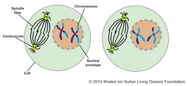 Figure 6-17 |
| Metaphase II | • Spindle fibers attach to the kinetochores.
• Chromosomes line up at the metaphase plate. |
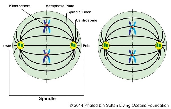 Figure 6-18 |
| Anaphase II | • The chromosomes split and each chromatid heads towards opposite poles.
• In preparation for division, the cell continues to elongate, forming a cleavage furrow. |
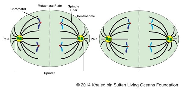 Figure 6-19 |
| Telophase II | • Chromosomes consist of one chromatid.
• The nuclear envelopes develop around each set of chromatids. • The spindle fibers disappear. |
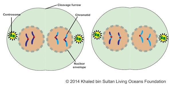 Figure 6-20 |
| Cytokinesis II | • The two daughter cells divide into four daughter cells. | 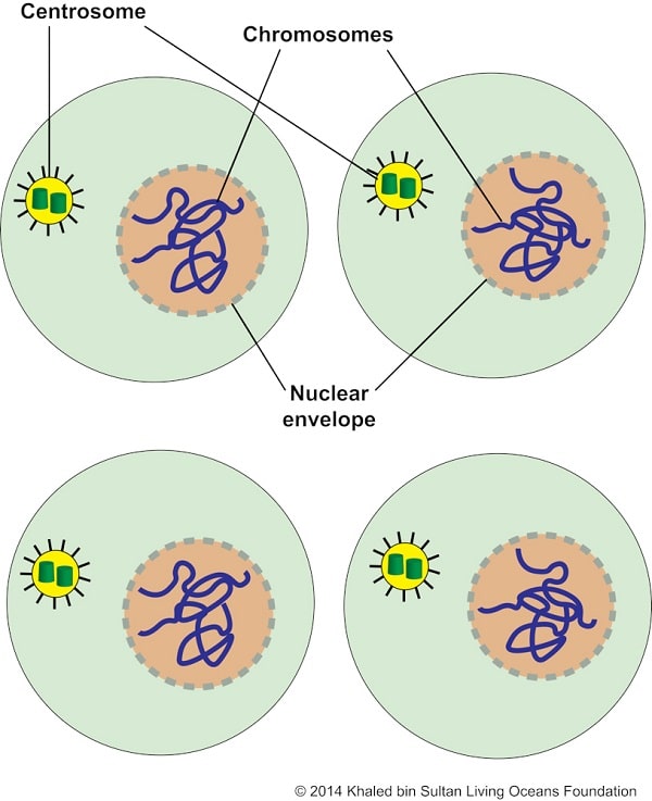 Figure 6-21 |

CORAL CYCLEs
Let’s recap the life and cell cycles of corals. Coral polyps become sexually mature and their cells undergo meiosis, producing gametes (egg and/or sperm). The gametes are fertilized, producing a zygote. The zygote continuously creates new cells by the process of mitosis, eventually forming a planula that settles and forms a coral polyp. As the coral grows, its cells continue to go through mitosis. When the coral polyp is sexually mature, it performs meiosis, starting the cycle all over again (figure 6-22).
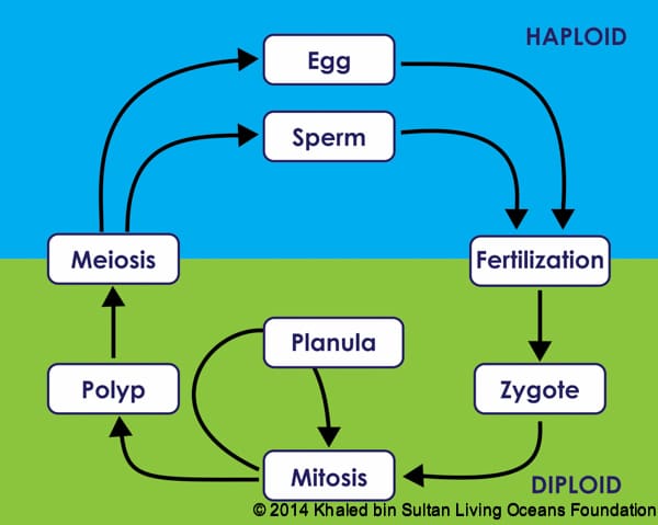
Figure 6-22. Coral polyp’s life cycle and cell cycles

APPENDIX
CHROMOSOME AND CELL TERMINOLOGY
Below is a review of some of the parts of a chromosome (figure 6-23) and a cell (figure 6-24).
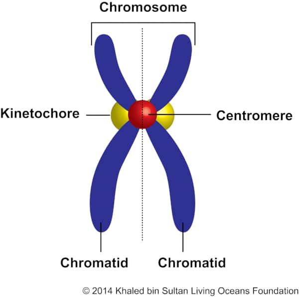
Centromere: The area on the chromosome where the kinetochores attach during cell division and that connects the chromatids together.
Chromosomes: Long strands of molecules called DNA.
Chromatid: When dividing the chromosome longitudinally, each half is considered a chromatid. They contain a double helix of DNA.
Kinetochore: A structure that forms on the centromere and is the point of attachment for microtubules during cell division.
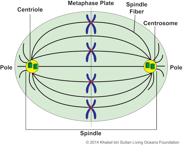
Centriole: A cylindrical organelle that consists of microtubules. They help to anchor the spindle fibers during cell division.
Centrosome: Replicates during interphase. It is part of the spindle and contains two centrioles. During cell division, they migrate to opposite poles and aid in pulling apart the chromosomes.
Metaphase plate: An imaginary vertical plane that divides the cell into two halves.
Microtubule: Miniature tubes that help to support the structure of the cell. It is like our skeleton and how it functions to support our body. They form spindle fibers during cell division.
Spindle fiber: Clusters of microtubules that help to move chromosomes during cell division.
Spindle: The structure that separates chromosomes during cell division. It contains the spindle fibers and centrosomes.

ATTRIBUTION
Figure 6-22. Adapted from Alternation of generations.svg: By Peter coxhead derivative work: Peter coxhead [Public domain], 20 February 2012 via Wikimedia Commons. http://commons.wikimedia.org/wiki/File%3AAlternation_of_generations_simpler.svg.
